세포 배양
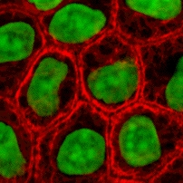
세포 배양(Cell culture)은 일반적으로 자연 환경 외부의 통제된 조건에서 세포가 성장하는 과정의 총칭이다. 세포는 살아있는 조직에서 분리된 후에는 신중하게 제어된 조건에서 후속적으로 유지되어야 한다. 이러한 조건은 각 세포 유형에 따라 다르지만 일반적으로 필수 영양소(아미노산, 탄수화물, 비타민, 무기질), 성장인자, 호르몬, 가스(이산화탄소, 산소)를 공급하는 기질 또는 배지가 있는 적합한 용기로 구성된다. 이는 물리화학적 환경(완충 용액, 삼투압, 온도)을 조절할 수 있게 설계되었다. 대부분의 세포는 단층(하나의 단일 세포 두께)으로 부착 배양을 형성하기 위해 표면 또는 인공 기질이 필요한 반면, 어떤 세포는 현탁액 배양으로 배지에 자유롭게 떠서 성장할 수 있다.[1] 대부분의 세포의 수명은 유전적으로 결정되지만 일부 세포 배양 세포는 최적의 조건이 제공되면 무한정 재생되도록 변환되기도 한다.


실제로 세포 배양이라는 용어는 식물 조직 배양, 진균 배양, 미생물 배양의 뜻보다 동물 세포에서 유래된 세포의 배양을 의미한다. 세포 배양의 역사적 발전과 방법은 조직 배양 및 장기 배양 과 밀접한 관련이 있다. 바이러스의 숙주인 세포와 함께 바이러스 배양도 관련이 있다.[2][3]
포유류 세포 배양의 개념
편집세포의 분리
편집세포는 여러 가지 방법으로 생체 외 배양을 위해 조직에서 분리한다. 세포는 현탁액으로 방출하기 위해 조직을 교반하기 전에 콜라겐 분해 효소, 트립신, 프로네이스와 같은 효소를 사용하여 세포 외 기질을 소화함으로써 고체 조직에서 분리한다.[4][5] 또는 조직 조각을 배지에 넣고 세포를 배양할 수 있다.
피험자로부터 직접 배양 된 세포를 1차 세포라고 한다. 일부 종양에서 유래한 것을 제외하고 대부분의 1차 세포는 수명이 제한되어 있다.
불멸 세포주는 무작위 돌연변이 또는 텔로머레이스 유전자의 인공적인 발현과 같은 의도적인 변형을 통해 무기한 증식하는 능력을 획득하였다. 특정 세포 유형을 대표하는 수 많은 세포주가 존재한다.
배양을 통한 세포 유지
편집대부분의 분리된 1차 세포는 생물학적 노화 과정을 거치고 일반적인 생존 능력(Hayflick 한계)을 유지하면서 특정 인구 수가 되면 분열을 멈춘다.

온도 및 가스 혼합물을 제외하고 배양 시스템에서 가장 일반적인 변화 요소는 세포 성장 배지이다. 성장 배지의 제조법은 수소 이온 농도 지수, 포도당 농도, 성장 인자 및 기타 영양소의 존재의 여부에 따라 다르다. 배지를 보충하는 데 사용되는 성장 인자는 종종 소태아혈청(FBS), 송아지 혈청, 말혈청, 돼지 혈청과 같은 동물 혈액의 혈청에서 파생된다. 이러한 혈액 유래 성분 성장 인자의 단점은 특히 의료 생명공학기술 응용 분야에서 바이러스 또는 프리온으로 배양물이 오염될 가능성이 있다는 것이다. 현재는 이러한 성분의 사용을 최소화하거나 제거하고 인간 혈소판 용해물(hPL)를 사용함으로 그 단점을 줄일 수 있다.[6] 이것은 FBS를 인간 세포와 함께 사용할 때 종간 오염에 대한 걱정을 없애준다. hPL은 FBS 또는 기타 동물 혈청을 직접 대체하는 안전하고 신뢰할 수 있는 대안으로 부상했다. 또한, 화학 배지를 사용하여 혈청 흔적(인간 또는 동물)을 제거할 수 있지만 이는 다른 세포 유형에서 항상 달성할 수 있는 것은 아니다. 대체 전략에는 미국, 호주 및 뉴질랜드와 같이 BSE/TSE 위험이 최소인 국가에서 동물 혈액을 조달하고[7] 세포 배양을 위해 전 동물 혈청 대신 혈청에서 추출한 정제된 영양 농축액을 사용하는 것이 포함된다.[8]
Plating 밀도(배양 배지 부피 당 세포 수)는 일부 세포 유형에서 중요한 역할을 한다. 예를 들어, Plating 밀도가 낮을수록 과립막 세포는 에스트로겐 생성을 나타내지만 Plating 밀도가 높으면 프로게스테론을 생성하는 테카 루테인 세포로 나타난다.[9]
세포는 현탁액 또는 부착 배양으로 성장할 수 있다.[10] 일부 세포는 혈류에 존재하는 세포와 같이 표면에 부착되지 않고 자연적으로 부유 상태로 존재한다. 부착 조건이 허용하는 것보다 더 높은 밀도로 성장할 수 있도록 현탁액 배양에서 생존할 수 있도록 변형된 세포주도 있다. 부착 세포는 부착 특성을 증가시키고 성장 및 분화에 필요한 기타 신호를 제공하기 위해 세포외 기질(콜라겐 및 라미닌) 성분으로 코팅될 수 있는 조직 배양 플라스틱 또는 미세 담체와 같은 표면이 필요하다. 고형 조직에서 유래한 대부분의 세포는 부착되어 있다. 부착 배양의 또 다른 유형은 2차원 배양 접시와 달리 3차원 환경에서 세포를 성장시키는 것을 포함하는 Organotypic 배양이다. 이 3D 배양 시스템은 생화학적 및 생리학적으로 생체 내 조직과 더 유사하지만 많은 요인(확산 등)으로 인해 유지 관리하기가 기술적으로 어렵다.[11]
세포 배양 기초 배지
편집생명과학에서 일상적으로 사용되는 세포 배양 배지이다.
- MEM
- DMEM
- RPMI 1640
- Ham's F-12
- IMDM
- Leibovitz L-15
- DMEM/F-12
세포 배양 배지의 구성 요소
편집| 요소 | 기능 |
|---|---|
| 탄소원(포도당/글루타민) | 에너지원 |
| 아미노산 | 단백질 제공 |
| 비타민 | 세포 생존 및 성장 촉진 |
| 적절한 농도의 염 | 세포 내에서 최적의 삼투압을 유지하고 효소 반응, 세포 부착 등의 보조 인자로 작용하는 필수 금속 이온을 제공하기 위한 이온의 등장성 혼합물 |
| 페놀 레드 염료 | 산·염기 지시약. 페놀 레드의 색상은 pH 7~7.4에서 주황색(혹은 빨간색)에서 산성에서는 노란색으로, 염기성에서는 자주색으로 바뀐다. |
| 중탄산염/HEPES 완충 용액 | 배지에서 균형 잡힌 수소 이온 농도를 유지하는 데 사용된다. |
전형적인 성장 조건
편집| 조건 | |
|---|---|
| 온도 | 37°C |
| 이산화탄소 | 5% |
| 상대 습도 | 95% |
세포주 교차 오염
편집세포주 교차 오염은 배양된 세포를 다루는 과학자에게 문제가 될 수 있다.[12] 연구에 따르면 15~20%의 시간에서 실험에 사용된 세포가 잘못 식별되었거나 다른 세포주로 오염된 것으로 나타났다.[13][14][15] 세포주 교차 오염 문제는 약물 스크리닝 연구에 일상적으로 사용되는 NCI-60의 세포주에서도 감지되었다.[16][17] ATCC(American Type Culture Collection), ECACC(European Collection of Cell Cultures), DSMZ(German Collection of Microorganisms and Cell Cultures)를 포함한 주요 세포주 저장소는 연구자로부터 잘못 식별된 세포주 제출을 받았다.[16][18] 이러한 오염은 세포 배양주를 사용하여 생산된 연구의 품질에 문제를 제기할 수 있다.[19] ATCC는 짧은 탠덤 반복( Short Tandem Repeat, STR), DNA 지문을 사용하여 오염되지 않은 세포주로 인증한다.[20]
세포주 교차 오염 문제를 해결하기 위해 연구자들은 세포주의 정체성을 확립하기 위해 초기 계대에서 세포주를 인증하는 것이 좋다. 세포주를 동결하기 전, 활성 배양 동안 2개월마다, 그리고 세포주를 사용하여 생성된 연구 데이터를 출판하기 전에 인증을 반복해야 한다. 동질효소 분석, 인간 림프구 항원(HLA) 유형, 염색체 분석, 핵형 분석, 형태학 및 STR 분석을 비롯한 많은 방법이 세포주를 식별하는 데 사용된다.[20]
중요한 세포주 교차 오염의 예중 하나는 헬라 세포주이다.
기타 기술적 문제
편집세포는 일반적으로 배양에서 계속 분열하기 때문에 일반적으로 사용 가능한 영역 또는 부피를 채우기 위해 성장한다. 이로 인해 몇 가지 문제가 발생할 수 있다.
- 성장 배지의 영양 고갈
- 성장 배지의 pH 변화
- 세포 자살/괴사 세포의 축적
- 세포 간 접촉은 세포 주기 정지를 자극하여 세포가 분열을 멈추게 하는 접촉 억제
- 세포 간 접촉에 따른 세포 분화
- 유전적 및 후성적 변화, 변형된 세포의 자연 선택으로 잠재적으로 분화가 감소하고 증식 능력이 증가된 비정상적 배양 적응 세포의 과성장[21]
배지의 선택은 영양 성분과 농도의 차이로 인해 세포 배양 실험 결과의 생리학적 관련성에 영향을 미칠 수 있다.[22][23] 영양소의 생리학적 수준을 더 잘 나타내는 배지를 사용하면 생체 외 연구의 생리적 관련성을 향상 시킬 수 있으며 최근에는 Plasmax[24] 및 인간 Plasma Like Medium(HPLM)[25]과 같은 배지 유형이 개발되었다.
배양 세포의 조작
편집배양 세포에서 수행되는 일반적인 조작 중에는 배지 변경, 세포 계대, 세포 형질 주입이 있다. 이들은 일반적으로 무균 기술에 의존하는 조직 배양 방법을 사용하여 수행된다. 무균 기술은 세균, 효모 또는 기타 세포주의 오염을 방지하는 것을 목표로 한다. 조작은 일반적으로 오염 미생물을 배제하기 위해 생물 안전 작업대 또는 층류 작업대에서 수행된다. 항생제(페니실린, 스트렙토마이신) 및 항진균제(암포테리신 B)도 배지에 첨가할 수 있다.
세포가 대사 과정을 거치면서 산이 생성되고 pH가 감소한다. 산·염기 지시약을 배지에 첨가하여 영양소 고갈을 측정한다.
부착 배양의 경우 흡인에 의해 배지를 직접 제거한 다음 교체할 수 있다.
세포의 Passage
편집계대 배양에는 소수의 세포를 새 용기로 옮기는 작업이 포함된다. 세포를 규칙적으로 분할하면 장기간 높은 세포 밀도와 관련된 노화를 방지하므로 세포를 더 오랜 시간 동안 배양할 수 있다. 현탁 배양은 더 많은 양의 신선한 배지에 희석된 몇 개의 세포를 포함하는 소량의 배양으로 쉽게 계대된다. 부착 배양의 경우 먼저 용기에서 세포를 분리해야 한다. 트립신/EDTA의 혼합물을 이용한다. 그런 다음 소수의 분리된 세포를 사용하여 새로운 용기에 옮겨 담는다. RAW 세포와 같은 일부 세포 배양은 고무 Scraper로 용기 표면에서 물리적으로 긁어낸다.
형질 전환 및 형질 도입
편집세포를 조작하는 또 다른 일반적인 방법은 형질 주입에 의한 외래 DNA의 도입을 포함한다. 이것은 세포가 관심 유전자를 발현하도록 하기 위해 수행된다. 최근에 RNA 간섭 구조의 형질 주입은 특정 유전자 및 단백질의 발현을 억제하기 위한 편리한 메커니즘으로 실현되었다. 또한 DNA는 형질 도입 또는 형질 전환이라고 하는 방법으로 바이러스를 사용하여 세포에 삽입될 수 있다. 바이러스는 기생체이기 때문에 정상적인 번식 과정의 일부이기 때문에 DNA를 세포에 도입하는 데 매우 적합하다.
확립된 인간 세포주 (미번역)
편집일반적인 세포주
편집인간 세포주
편집- H295R (부신피질암)
- DU145 (전립선암)
- HeLa ( 자궁경부암 )
- KBM-7 (만성 골수성 백혈병)
- LNCaP (전립선암)
- MCF-7 (유방암)
- MDA-MB-468 (유방암)
- PC3 (전립선암)
- SaOS-2 (골종양)
- SH-SY5Y (신경모세포종, 다발성 골수종)
- T-47D (유방암)
- THP-1 (급성 골수성 백혈병)
- U87 (교모세포종)
영장류 세포주
편집쥐 세포주
편집- MC3T3 (배아 Calvarium)
쥐 종양 세포주
편집식물 세포주
편집기타 종 세포주
편집세포주 목록
편집| 세포주 | 의미 | 생물체 | 조직 출처 | 형태 | 링크 |
|---|---|---|---|---|---|
| 3T3-L1 | 3-day Transfer, Inoculum 3x10⁵ cells | 쥐 | Embryo | 섬유아세포 | ECACC Cellosaurus |
| 4T1 | 쥐 | Mammary gland | ATCC Cellosaurus | ||
| 1321N1 | 인간 | 뇌 | Astrocytoma | ECACC Cellosaurus | |
| 9L | 쥐 | 뇌 | Glioblastoma | ECACC Cellosaurus | |
| A172 | 인간 | 뇌 | Glioblastoma | ECACC Cellosaurus | |
| A20 | 쥐 | B lymphoma | B lymphocyte | Cellosaurus | |
| A253 | 인간 | Submandibular duct | Head and neck carcinoma | ATCC Cellosaurus | |
| A2780 | 인간 | 난소 | Ovarian carcinoma | ECACC Cellosaurus | |
| A2780ADR | 인간 | 난소 | Adriamycin-resistant derivative of A2780 | ECACC Cellosaurus | |
| A2780cis | 인간 | 난소 | Cisplatin-resistant derivative of A2780 | ECACC Cellosaurus | |
| A431 | 인간 | Skin epithelium | Squamous cell carcinoma | ECACC Cellosaurus | |
| A549 | 인간 | 폐 | Lung carcinoma | ECACC Cellosaurus | |
| AB9 | 제브라피쉬 | Fin | 섬유아세포 | ATCC Cellosaurus | |
| AHL-1 | Armenian Hamster Lung-1 | 햄스터 | 폐 | ECACC Archived 2021년 11월 24일 - 웨이백 머신 Cellosaurus | |
| ALC | 쥐 | 골수 | Stroma | PMID 2435412[26] Cellosaurus | |
| B16 | 쥐 | Melanoma | ECACC Archived 2021년 11월 24일 - 웨이백 머신 Cellosaurus | ||
| B35 | 쥐 | Neuroblastoma | ATCC Cellosaurus | ||
| BCP-1 | 인간 | 말초 혈액 단핵세포(PBMC) | HIV+ primary effusion lymphoma | ATCC Cellosaurus | |
| BEAS-2B | Bronchial epithelium + Adenovirus 12-SV40 virus hybrid (Ad12SV40) | 인간 | 폐 | Epithelial | ECACC Cellosaurus |
| bEnd.3 | Brain Endothelial 3 | 쥐 | 뇌, 대뇌 피질 | Endothelium | Cellosaurus |
| BHK-21 | Baby Hamster Kidney-21 | 햄스터 | 신장 | 섬유아세포 | ECACC Archived 2021년 11월 24일 - 웨이백 머신 Cellosaurus |
| BOSC23 | Packaging cell line derived from HEK 293 | 인간 | Kidney (embryonic) | Epithelium | Cellosaurus |
| BT-20 | Breast Tumor-20 | 인간 | Breast epithelium | Breast carcinoma | ATCC Cellosaurus |
| BxPC-3 | Biopsy xenograft of Pancreatic Carcinoma line 3 | 인간 | Pancreatic adenocarcinoma | Epithelial | ECACC Cellosaurus |
| C2C12 | 쥐 | Myoblast | ECACC Cellosaurus | ||
| C3H-10T1/2 | 쥐 | Embryonic mesenchymal cell line | ECACC Cellosaurus | ||
| C6 | 쥐 | Brain astrocyte | Glioma | ECACC Cellosaurus | |
| C6/36 | 흰줄숲모기 | Larval tissue | ECACC Cellosaurus | ||
| Caco-2 | 인간 | 대장 | Colorectal carcinoma | ECACC Cellosaurus | |
| Cal-27 | 인간 | 혀 | Squamous cell carcinoma | ATCC Cellosaurus | |
| Calu-3 | 인간 | 폐 | Adenocarcinoma | ATCC Cellosaurus | |
| CGR8 | 쥐 | Embryonic stem cells | ECACC Cellosaurus | ||
| CHO | Chinese Hamster Ovary | 햄스터 | 난소 | Epithelium | ECACC Archived 2021년 10월 29일 - 웨이백 머신 Cellosaurus |
| CML T1 | Chronic myeloid leukemia T lymphocyte 1 | 인간 | CML acute phase | T cell leukemia | DSMZ Cellosaurus |
| CMT12 | Canine Mammary Tumor 12 | 개 | Mammary gland | Epithelium | Cellosaurus |
| COR-L23 | 인간 | 폐 | Lung carcinoma | ECACC Cellosaurus | |
| COR-L23/5010 | 인간 | 폐 | Lung carcinoma | ECACC Cellosaurus | |
| COR-L23/CPR | 인간 | 폐 | Lung carcinoma | ECACC Cellosaurus | |
| COR-L23/R23- | 인간 | 폐 | Lung carcinoma | ECACC Cellosaurus | |
| COS-7 | Cercopithecus aethiops, origin-defective SV-40 | Old World monkey - Cercopithecus aethiops (Chlorocebus) | 신장 | 섬유아세포 | ECACC Cellosaurus |
| COV-434 | 인간 | 난소 | Ovarian granulosa cell carcinoma | PMID 8436435[27] ECACC Cellosaurus | |
| CT26 | 쥐 | 대장 | Colorectal carcinoma | Cellosaurus | |
| D17 | 개 | Lung metastasis | Osteosarcoma | ATCC Cellosaurus | |
| DAOY | 인간 | 뇌 | Medulloblastoma | ATCC Cellosaurus | |
| DH82 | 개 | Histiocytosis | Monocyte/macrophage | ECACC Cellosaurus | |
| DU145 | 인간 | Androgen insensitive prostate carcinoma | ATCC Cellosaurus | ||
| DuCaP | Dura mater cancer of the Prostate | 인간 | Metastatic prostate carcinoma | Epithelial | PMID 11317521[28] Cellosaurus |
| E14Tg2a | 쥐 | Embryonic stem cells | ECACC Cellosaurus | ||
| EL4 | 쥐 | T cell leukemia | ECACC Cellosaurus | ||
| EM-2 | 인간 | CML blast crisis | Ph+ CML line | DSMZ Cellosaurus | |
| EM-3 | 인간 | CML blast crisis | Ph+ CML line | DSMZ Cellosaurus | |
| EMT6/AR1 | 쥐 | Mammary gland | Epithelial-like | ECACC Cellosaurus | |
| EMT6/AR10.0 | 쥐 | Mammary gland | Epithelial-like | ECACC Cellosaurus | |
| FM3 | 인간 | Lymph node metastasis | Melanoma | ECACC Cellosaurus | |
| GL261 | Glioma 261 | 쥐 | 뇌 | Glioma | Cellosaurus |
| H1299 | 인간 | 폐 | Lung carcinoma | ATCC Cellosaurus | |
| HaCaT | 인간 | 피부 | Keratinocyte | CLS Archived 2021년 11월 24일 - 웨이백 머신 Cellosaurus | |
| HCA2 | 인간 | Colon | Adenocarcinoma | ECACC Cellosaurus | |
| HEK 293 | Human Embryonic Kidney 293 | 인간 | Kidney (embryonic) | Epithelium | ECACC Cellosaurus |
| HEK 293T | HEK 293 derivative | 인간 | Kidney (embryonic) | Epithelium | ECACC Cellosaurus |
| HeLa | Henrietta Lacks | 인간 | Cervix epithelium | Cervical carcinoma | ECACC Cellosaurus |
| Hepa1c1c7 | Clone 7 of clone 1 hepatoma line 1 | 쥐 | Hepatoma | Epithelial | ECACC Cellosaurus |
| Hep G2 | 인간 | 간 | Hepatoblastoma | ECACC Cellosaurus | |
| High Five | Insect (moth) - Trichoplusia ni | Ovary | Cellosaurus | ||
| HL-60 | Human Leukemia-60 | 인간 | 혈액 | Myeloblast | ECACC Cellosaurus |
| HT-1080 | 인간 | Fibrosarcoma | ECACC Cellosaurus | ||
| HT-29 | 인간 | Colon epithelium | Adenocarcinoma | ECACC Cellosaurus | |
| J558L | 쥐 | Myeloma | B lymphocyte cell | ECACC Cellosaurus | |
| Jurkat | 인간 | White blood cells | T cell leukemia | ECACC Cellosaurus | |
| JY | 인간 | Lymphoblastoid | EBV-transformed B cell | ECACC Cellosaurus | |
| K562 | 인간 | Lymphoblastoid | CML blast crisis | ECACC Cellosaurus | |
| KBM-7 | 인간 | Lymphoblastoid | CML blast crisis | Cellosaurus | |
| KCL-22 | 인간 | Lymphoblastoid | CML | DSMZ Cellosaurus | |
| KG1 | 인간 | Lymphoblastoid | AML | ECACC Cellosaurus | |
| Ku812 | 인간 | Lymphoblastoid | Erythroleukemia | ECACC Cellosaurus | |
| KYO-1 | Kyoto-1 | 인간 | Lymphoblastoid | CML | DSMZ Cellosaurus |
| L1210 | 쥐 | Lymphocytic leukemia | Ascitic fluid | ECACC Cellosaurus | |
| L243 | 쥐 | Hybridoma | Secretes L243 mAb (against HLA-DR) | ATCC Cellosaurus | |
| LNCaP | Lymph Node Cancer of the Prostate | 인간 | Prostatic adenocarcinoma | Epithelial | ECACC Cellosaurus |
| MA-104 | Microbiological Associates-104 | 아프리카 녹색 원숭이 | Kidney | Epithelial | Cellosaurus |
| MA2.1 | 쥐 | Hybridoma | Secretes MA2.1 mAb (against HLA-A2 and HLA-B17) | ATCC Cellosaurus | |
| Ma-Mel 1, 2, 3....48 | 인간 | Skin | A range of melanoma cell lines | ECACC Archived 2021년 11월 24일 - 웨이백 머신 Cellosaurus | |
| MC-38 | Mouse Colon-38 | 쥐 | Colon | Adenocarcinoma | Cellosaurus |
| MCF-7 | Michigan Cancer Foundation-7 | 인간 | Breast | Invasive breast ductal carcinoma ER+, PR+ | ECACC Cellosaurus |
| MCF-10A | Michigan Cancer Foundation-10A | 인간 | Breast epithelium | ATCC Cellosaurus | |
| MDA-MB-157 | M.D. Anderson - Metastatic Breast-157 | 인간 | Pleural effusion metastasis | Breast carcinoma | ECACC Cellosaurus |
| MDA-MB-231 | M.D. Anderson - Metastatic Breast-231 | 인간 | Pleural effusion metastasis | Breast carcinoma | ECACC Cellosaurus |
| MDA-MB-361 | M.D. Anderson - Metastatic Breast-361 | 인간 | Melanoma (contaminated by M14) | ECACC Cellosaurus | |
| MDA-MB-468 | M.D. Anderson - Metastatic Breast-468 | 인간 | Pleural effusion metastasis | Breast carcinoma | ATCC Cellosaurus |
| MDCK II | Madin Darby Canine Kidney II | 개 | Kidney | Epithelium | ECACC Cellosaurus |
| MG63 | 인간 | Bone | Osteosarcoma | ECACC Cellosaurus | |
| MIA PaCa-2 | 인간 | Prostate | Pancreatic Carcinoma | ATCC Cellosaurus | |
| MOR/0.2R | 인간 | Lung | Lung carcinoma | ECACC Cellosaurus | |
| Mono-Mac-6 | 인간 | White blood cells | Myeloid metaplasic AML | DSMZ Cellosaurus | |
| MRC-5 | Medical Research Council cell strain 5 | 인간 | Lung (fetal) | 섬유아세포 | ECACC Archived 2021년 11월 24일 - 웨이백 머신 Cellosaurus |
| MTD-1A | 쥐 | Epithelium | Cellosaurus | ||
| MyEnd | Myocardial Endothelial | 쥐 | Endothelium | Cellosaurus | |
| NCI-H69 | 인간 | Lung | Lung carcinoma | ECACC Cellosaurus | |
| NCI-H69/CPR | 인간 | Lung | Lung carcinoma | ECACC Cellosaurus | |
| NCI-H69/LX10 | 인간 | Lung | Lung carcinoma | ECACC Cellosaurus | |
| NCI-H69/LX20 | 인간 | Lung | Lung carcinoma | ECACC Cellosaurus | |
| NCI-H69/LX4 | 인간 | Lung | Lung carcinoma | ECACC Cellosaurus | |
| Neuro-2a | 쥐 | Nerve/neuroblastoma | Neuronal stem cells | ECACC Cellosaurus | |
| NIH-3T3 | NIH, 3-day transfer, inoculum 3x10⁵ cells | 쥐 | Embryo | 섬유아세포 | ECACC Cellosaurus |
| NALM-1 | 인간 | Peripheral blood | Blast-crisis CML | ATCC Cellosaurus | |
| NK-92 | 인간 | Leukemia/lymphoma | ATCC Cellosaurus | ||
| NTERA-2 | 인간 | Lung metastasis | Embryonal carcinoma | ECACC Cellosaurus | |
| NW-145 | 인간 | Skin | Melanoma | ESTDAB Archived 2011년 11월 16일 - 웨이백 머신 Cellosaurus | |
| OK | Opossum Kidney | Virginia opossum - Didelphis virginiana | Kidney | ECACC Cellosaurus | |
| OPCN / OPCT cell lines | 인간 | Prostate | Range of prostate tumour lines | Cellosaurus | |
| P3X63Ag8 | 쥐 | Myeloma | ECACC Cellosaurus | ||
| PANC-1 | 인간 | Duct | Epithelioid Carcinoma | ATCC Cellosaurus | |
| PC12 | 쥐 | Adrenal medulla | Pheochromocytoma | ECACC Cellosaurus | |
| PC-3 | Prostate Cancer-3 | 인간 | Bone metastasis | Prostate carcinoma | ECACC Cellosaurus |
| Peer | 인간 | T cell leukemia | DSMZ Cellosaurus | ||
| PNT1A | 인간 | Prostate | SV40-transformed tumour line | ECACC Cellosaurus | |
| PNT2 | 인간 | Prostate | SV40-transformed tumour line | ECACC Cellosaurus | |
| Pt K2 | The second cell line derived from Potorous tridactylis | Long-nosed potoroo - Potorous tridactylus | Kidney | Epithelial | ECACC Cellosaurus |
| Raji | 인간 | B lymphoma | Lymphoblast-like | ECACC Cellosaurus | |
| RBL-1 | Rat Basophilic Leukemia-1 | 쥐 | Leukemia | Basophil cell | ECACC Cellosaurus |
| RenCa | Renal Carcinoma | Mouse | Kidney | Renal carcinoma | ATCC Cellosaurus |
| RIN-5F | 쥐 | Pancreas | ECACC Cellosaurus | ||
| RMA-S | 쥐 | T cell tumour | Cellosaurus | ||
| S2 | Schneider 2 | Insect - Drosophila melanogaster | Late stage (20–24 hours old) embryos | ATCC Cellosaurus | |
| SaOS-2 | Sarcoma OSteogenic-2 | 인간 | Bone | Osteosarcoma | ECACC Cellosaurus |
| Sf21 | Spodoptera frugiperda 21 | Insect (moth) - Spodoptera frugiperda | Ovary | ECACC Cellosaurus | |
| Sf9 | Spodoptera frugiperda 9 | Insect (moth) - Spodoptera frugiperda | Ovary | ECACC Cellosaurus | |
| SH-SY5Y | 인간 | Bone marrow metastasis | Neuroblastoma | ECACC Cellosaurus | |
| SiHa | 인간 | Cervix epithelium | Cervical carcinoma | ATCC Cellosaurus | |
| SK-BR-3 | Sloan-Kettering Breast cancer 3 | 인간 | Breast | Breast carcinoma | DSMZ Cellosaurus |
| SK-OV-3 | Sloan-Kettering Ovarian cancer 3 | 인간 | Ovary | Ovarian carcinoma | ECACC Cellosaurus |
| SK-N-SH | 인간 | Brain | Epithelial | ATCC Cellosaurus | |
| T2 | 인간 | T cell leukemia/B cell line hybridoma | ATCC Cellosaurus | ||
| T-47D | 인간 | Breast | Breast ductal carcinoma | ECACC Cellosaurus | |
| T84 | 인간 | Lung metastasis | Colorectal carcinoma | ECACC Cellosaurus | |
| T98G | 인간 | Glioblastoma-astrocytoma | Epithelium | ECACC Cellosaurus | |
| THP-1 | 인간 | Monocyte | Acute monocytic leukemia | ECACC Cellosaurus | |
| U2OS | 인간 | Osteosarcoma | Epithelial | ECACC Cellosaurus | |
| U373 | 인간 | Glioblastoma-astrocytoma | Epithelium | ECACC Archived 2021년 11월 24일 - 웨이백 머신 Cellosaurus | |
| U87 | 인간 | Glioblastoma-astrocytoma | Epithelial-like | ECACC Cellosaurus | |
| U937 | 인간 | Leukemic monocytic lymphoma | ECACC Cellosaurus | ||
| VCaP | Vertebral Cancer of the Prostate | 인간 | Vertebra metastasis | Prostate carcinoma | ECACC Cellosaurus |
| Vero | From Esperanto: verda (green, for green monkey) reno (kidney) | African green monkey - Chlorocebus sabaeus | Kidney epithelium | ECACC Cellosaurus | |
| VG-1 | 인간 | Primary effusion lymphoma | Cellosaurus | ||
| WM39 | 인간 | Skin | Melanoma | ESTDAB Archived 2021년 4월 27일 - 웨이백 머신 Cellosaurus | |
| WT-49 | 인간 | Lymphoblastoid | ECACC Cellosaurus | ||
| YAC-1 | 쥐 | Lymphoma | ECACC Cellosaurus | ||
| YAR | 인간 | Lymphoblastoid | EBV-transformed B cell | 인간 Immunology[29] ECACC Cellosaurus |
같이 보기
편집- 생물학적 영생
- 세포 배양 분석
- 전기적 세포-기질 임피던스 감지
- 오염된 세포주 목록
- NCI-60 세포주의 목록
- 유방암 세포주 목록
- 미세생리계
각주
편집- ↑ Harris, AR; Peter, L; Bellis, J; Baum, B; Kabla, AJ; Charras, GT (2012년 10월 9일). “Characterizing the mechanics of cultured cell monolayers.”. 《Proceedings of the National Academy of Sciences of the United States of America》 109 (41): 16449–54. doi:10.1073/pnas.1213301109. PMC 3478631. PMID 22991459.
- ↑ “Some landmarks in the development of tissue and cell culture”. 2006년 4월 19일에 확인함.
- ↑ “Cell Culture”. 2006년 4월 19일에 확인함.
- ↑ “Methods for isolating atrial cells from large mammals and humans”. 《Journal of Molecular and Cellular Cardiology》 86: 187–98. September 2015. doi:10.1016/j.yjmcc.2015.07.006. PMID 26186893.
- ↑ “Methods in cardiomyocyte isolation, culture, and gene transfer”. 《Journal of Molecular and Cellular Cardiology》 51 (3): 288–98. September 2011. doi:10.1016/j.yjmcc.2011.06.012. PMC 3164875. PMID 21723873.
- ↑ Hemeda, H., Giebel, B., Wagner, W. (16Feb2014) Evaluation of human platelet lysate versus fetal bovine serum for culture of mesenchymal stromal cells Cytotherapy p170-180 issue 2 doi.10.1016
- ↑ “Post - Blog | Boval BioSolutions, LLC”. bovalco.com. 2014년 9월 10일에 원본 문서에서 보존된 문서. 2014년 12월 2일에 확인함.
- ↑ “LipiMAX purified lipoprotein solution from bovine serum”. 《Selborne Biological Services》. 2006. 2012년 6월 8일에 원본 문서에서 보존된 문서. 2010년 2월 2일에 확인함.
- ↑ “Cell plating density alters the ratio of estrogenic to progestagenic enzyme gene expression in cultured granulosa cells”. 《Fertility and Sterility》 93 (6): 2050–5. April 2010. doi:10.1016/j.fertnstert.2009.01.151. PMID 19324349.
- ↑ Jaccard, N; Macown, RJ; Super, A; Griffin, LD; Veraitch, FS; Szita, N (October 2014). “Automated and online characterization of adherent cell culture growth in a microfabricated bioreactor.”. 《Journal of Laboratory Automation》 19 (5): 437–43. doi:10.1177/2211068214529288. PMC 4230958. PMID 24692228.
- ↑ “Organotypic brain slice cultures: A review”. 《Neuroscience》 305: 86–98. October 2015. doi:10.1016/j.neuroscience.2015.07.086. PMC 4699268. PMID 26254240.
- ↑ “Line of attack”. 《Science》 347 (6225): 938–40. February 2015. Bibcode:2015Sci...347..938N. doi:10.1126/science.347.6225.938. PMID 25722392.
- ↑ “False human hematopoietic cell lines: cross-contaminations and misinterpretations”. 《Leukemia》 13 (10): 1601–7. October 1999. doi:10.1038/sj.leu.2401510. PMID 10516762.
- ↑ “Cross-contamination: HS-Sultan is not a myeloma but a Burkitt lymphoma cell line”. 《Blood》 98 (12): 3495–6. December 2001. doi:10.1182/blood.V98.12.3495. PMID 11732505.
- ↑ “Identity tests: determination of cell line cross-contamination”. 《Cytotechnology》 51 (2): 45–50. June 2006. doi:10.1007/s10616-006-9013-8. PMC 3449683. PMID 19002894.
- ↑ 가 나 “Cell biology. Cases of mistaken identity”. 《Science》 315 (5814): 928–31. February 2007. doi:10.1126/science.315.5814.928. PMID 17303729.
- ↑ “A case study in misidentification of cancer cell lines: MCF-7/AdrR cells (re-designated NCI/ADR-RES) are derived from OVCAR-8 human ovarian carcinoma cells”. 《Cancer Letters》 245 (1–2): 350–2. January 2007. doi:10.1016/j.canlet.2006.01.013. PMID 16504380.
- ↑ “Widespread intraspecies cross-contamination of human tumor cell lines arising at source”. 《International Journal of Cancer》 83 (4): 555–63. November 1999. doi:10.1002/(SICI)1097-0215(19991112)83:4<555::AID-IJC19>3.0.CO;2-2. PMID 10508494.
- ↑ “HeLa cells 50 years on: the good, the bad and the ugly”. 《Nature Reviews. Cancer》 2 (4): 315–9. April 2002. doi:10.1038/nrc775. PMID 12001993.
- ↑ 가 나 Dunham, J.H.; Guthmiller, P. (2008). “Doing good science: Authenticating cell line identity” (PDF). 《Cell Notes》 22: 15–17. 2008년 10월 28일에 원본 문서 (PDF)에서 보존된 문서. 2008년 10월 28일에 확인함.
- ↑ “Genetic and epigenetic instability in human pluripotent stem cells”. 《Human Reproduction Update》 19 (2): 187–205. 2012. doi:10.1093/humupd/dms048. PMID 23223511.
- ↑ Lagziel S; Gottlieb E; Shlomi T (2020). “Mind your media”. 《Nature Metabolism》 2 (12): 1369–1372. doi:10.1038/s42255-020-00299-y. PMID 33046912.
- ↑ Lagziel S; Lee WD; Shlomi T (2019). “Inferring cancer dependencies on metabolic genes from large-scale genetic screens.”
|url=값 확인 필요 (도움말). 《BMC Biol》 17 (1): 37. doi:10.1186/s12915-019-0654-4. PMC 6489231. PMID 31039782. - ↑ Vande Voorde J; Ackermann T; Pfetzer N; Sumpton D; Mackay G; Kalna G; 외. (2019). “Improving the metabolic fidelity of cancer models with a physiological cell culture medium.”
|url=값 확인 필요 (도움말). 《Sci Adv》 5 (1): eaau7314. Bibcode:2019SciA....5.7314V. doi:10.1126/sciadv.aau7314. PMC 6314821. PMID 30613774. - ↑ Cantor JR; Abu-Remaileh M; Kanarek N; Freinkman E; Gao X; Louissaint A; 외. (2017). “Physiologic Medium Rewires Cellular Metabolism and Reveals Uric Acid as an Endogenous Inhibitor of UMP Synthase.”
|url=값 확인 필요 (도움말). 《Cell》 169 (2): 258–272.e17. doi:10.1016/j.cell.2017.03.023. PMC 5421364. PMID 28388410. - ↑ “A single bone marrow-derived stromal cell type supports the in vitro growth of early lymphoid and myeloid cells”. 《Cell》 48 (6): 997–1007. March 1987. doi:10.1016/0092-8674(87)90708-2. PMID 2435412.
- ↑ “Establishment and characterization of 7 ovarian carcinoma cell lines and one granulosa tumor cell line: growth features and cytogenetics”. 《International Journal of Cancer》 53 (4): 613–20. February 1993. doi:10.1002/ijc.2910530415. PMID 8436435.
- ↑ “Establishment and characterization of a new human prostatic cancer cell line: DuCaP”. 《In Vivo》 15 (2): 157–62. 2001. PMID 11317521.
- ↑ “Promiscuous T-cell recognition of a rubella capsid protein epitope restricted by DRB1*0403 and DRB1*0901 molecules sharing an HLA DR supertype”. 《Human Immunology》 59 (3): 149–57. March 1998. doi:10.1016/S0198-8859(98)00006-8. PMID 9548074.