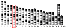
Peroxisome proliferator-activated receptor gamma coactivator 1-alpha (PGC-1α) is a protein that in humans is encoded by the PPARGC1A gene.[4] PPARGC1A is also known as human accelerated region 20 (HAR20). It may, therefore, have played a key role in differentiating humans from apes.[5]
It has been suggested that Pparg coactivator 1 alpha be merged into this article. (Discuss) Proposed since March 2024. |
PGC-1α is the master regulator of mitochondrial biogenesis.[6][7][8] PGC-1α is also the primary regulator of liver gluconeogenesis, inducing increased gene expression for gluconeogenesis.[9]
Function
editPGC-1α is a gene that contains two promoters, and has 4 alternative splicings. PGC-1α is a transcriptional coactivator that regulates the genes involved in energy metabolism. It is the master regulator of mitochondrial biogenesis.[6][7][8] This protein interacts with the nuclear receptor PPAR-γ, which permits the interaction of this protein with multiple transcription factors. This protein can interact with, and regulate the activity of, cAMP response element-binding protein (CREB) and nuclear respiratory factors (NRFs) [citation needed]. PGC-1α provides a direct link between external physiological stimuli and the regulation of mitochondrial biogenesis, and is a major factor causing slow-twitch rather than fast-twitch muscle fiber types.[10]
Endurance exercise has been shown to activate the PGC-1α gene in human skeletal muscle.[11] Exercise-induced PGC-1α in skeletal muscle increases autophagy[12][13] and unfolded protein response.[14]
PGC-1α protein may also be involved in controlling blood pressure, regulating cellular cholesterol homeostasis, and the development of obesity.[15]
Regulation
editPGC-1α is thought to be a master integrator of external signals. It is known to be activated by a host of factors, including:
- Reactive oxygen species and reactive nitrogen species, both formed endogenously in the cell as by-products of metabolism but upregulated during times of cellular stress.
- Fasting can also increase gluconeogenic gene expression, including hepatic PGC-1α.[16][17]
- It is strongly induced by cold exposure, linking this environmental stimulus to adaptive thermogenesis.[18]
- It is induced by endurance exercise[11] and recent research has shown that PGC-1α determines lactate metabolism, thus preventing high lactate levels in endurance athletes and making lactate as an energy source more efficient.[19]
- cAMP response element-binding (CREB) proteins, activated by an increase in cAMP following external cellular signals.
- Protein kinase B (Akt) is thought to downregulate PGC-1α, but upregulate its downstream effectors, NRF1 and NRF2. Akt itself is activated by PIP3, often upregulated by PI3K after G protein signals. The Akt family is also known to activate pro-survival signals as well as metabolic activation.
- SIRT1 binds and activates PGC-1α through deacetylation inducing gluconeogenesis without affecting mitochondrial biogenesis.[20]
PGC-1α has been shown to exert positive feedback circuits on some of its upstream regulators:
- PGC-1α increases Akt (PKB) and Phospho-Akt (Ser 473 and Thr 308) levels in muscle.[21]
- PGC-1α leads to calcineurin activation.[22]
Akt and calcineurin are both activators of NF-kappa-B (p65).[23][24] Through their activation, PGC-1α seems to activate NF-kappa-B. Increased activity of NF-kappa-B in muscle has recently been demonstrated following induction of PGC-1α.[25] The finding seems to be controversial. Other groups found that PGC-1s inhibit NF-kappa-B activity.[26] The effect was demonstrated for PGC-1 alpha and beta.
PGC-1α has also been shown to drive NAD biosynthesis to play a large role in renal protection in acute kidney injury.[27]
Clinical significance
editRecently PPARGC1A has been implicated as a potential therapy for Parkinson's disease conferring protective effects on mitochondrial metabolism.[28]
Moreover, brain-specific isoforms of PGC-1alpha have recently been identified which are likely to play a role in other neurodegenerative disorders such as Huntington's disease and amyotrophic lateral sclerosis.[29][30]
Massage therapy appears to increase the amount of PGC-1α, which leads to the production of new mitochondria.[31][32][33]
PGC-1α and beta has furthermore been implicated in polarization to anti-inflammatory M2 macrophages by interaction with PPAR-γ[34] with upstream activation of STAT6.[35] An independent study confirmed the effect of PGC-1 on polarisation of macrophages towards M2 via STAT6/PPAR gamma and furthermore demonstrated that PGC-1 inhibits proinflammatory cytokine production.[36]
PGC-1α has been recently proposed to be responsible for β-aminoisobutyric acid secretion by exercising muscles.[37] The effect of β-aminoisobutyric acid in white fat includes the activation of thermogenic genes that prompt the browning of white adipose tissue and the consequent increase of background metabolism. Hence, the β-aminoisobutyric acid could act as a messenger molecule of PGC-1α and explain the effects of PGC-1α increase in other tissues such as white fat.
PGC-1α increases BNP expression by coactivating ERRα and / or AP1. Subsequently, BNP induces a chemokine cocktail in muscle fibers and activates macrophages in a local paracrine manner, which can then contribute to enhancing the repair and regeneration potential of trained muscles.
Most studies reporting effects of PGC-1α on physiological functions have used mouse models in which the PGC-1α gene is either knocked out or overexpressed from conception. However, some of the proposed effects of PGC-1α have been questioned by studies using inducible knockout technology to remove the PGC-1α gene only in adult mice. For example, two independent studies have shown that adult expression of PGC-1α is not required for improved mitochondrial function after exercise training.[38][39] This suggests that some of the reported effects of PGC-1α are likely to occur only in the developmental stage.
Interactions
editPPARGC1A has been shown to interact with:
- CREB-binding protein[40]
- Estrogen-related receptor alpha (ERRα),[41] estrogen-related receptor beta (ERR-β), estrogen-related receptor gamma (ERR-γ).
- Farnesoid X receptor[42]
- FBXW7[43]
- MED1,[44] MED12,[44] MED14,[44] MED17,[44]
- NRF1[45]
- Peroxisome proliferator-activated receptor gamma[40][44]
- Retinoid X receptor alpha[46]
- Thyroid hormone receptor beta[47]
ERRα and PGC-1α are coactivators of both glucokinase (GK) and SIRT3, binding to an ERRE element in the GK and SIRT3 promoters.[citation needed]
See also
editReferences
editFurther reading
edit- Knutti D, Kralli A (2001). "PGC-1, a versatile coactivator". Trends Endocrinol. Metab. 12 (8): 360–5. doi:10.1016/S1043-2760(01)00457-X. PMID 11551810. S2CID 24230985.
- Puigserver P, Spiegelman BM (2003). "Peroxisome proliferator-activated receptor-gamma coactivator 1 alpha (PGC-1 alpha): transcriptional coactivator and metabolic regulator". Endocr. Rev. 24 (1): 78–90. doi:10.1210/er.2002-0012. PMID 12588810.
- Soyal S, Krempler F, Oberkofler H, Patsch W (2007). "PGC-1alpha: a potent transcriptional cofactor involved in the pathogenesis of type 2 diabetes". Diabetologia. 49 (7): 1477–88. doi:10.1007/s00125-006-0268-6. PMID 16752166.
- Handschin C, Spiegelman BM (2007). "Peroxisome proliferator-activated receptor gamma coactivator 1 coactivators, energy homeostasis, and metabolism". Endocr. Rev. 27 (7): 728–35. doi:10.1210/er.2006-0037. PMID 17018837.
External links
edit- PPARGC1A protein, human at the U.S. National Library of Medicine Medical Subject Headings (MeSH)
- NURSA C110
- FactorBook PGC1A
- Overview of all the structural information available in the PDB for UniProt: Q9UBK2 (Peroxisome proliferator-activated receptor gamma coactivator 1-alpha) at the PDBe-KB.
This article incorporates text from the United States National Library of Medicine, which is in the public domain.


